
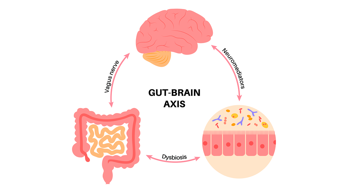
Imagine a constant dialogue inside our body: the exchange of information taking place between the gut and the brain. When we are anxious our "stomach ties itself in knots", stress can give us diarrhoea, and some of our decisions we make "on a hunch", from the gut — these expressions were not born by chance. Our gut is not merely a "digestive tube" but the home of a complex nervous network, often called the second brain[1]. The human gut contains some 200–600 million nerve cells — roughly as many as the spinal cord[1] — forming its own independent nervous system (the enteric nervous system, ENS). This neural "network city" communicates continuously with the central nervous system, among other things via the vagus nerve (nervus vagus)[2]. At the same time, another invisible "organ" lives with us in our gut: our microbiome, a community of many trillions of bacteria, viruses and other microbes. This microbial ecosystem produces an astonishing number of chemical signals and substances that can affect our body and even the workings of our brain[3]. The system of connections between the nerve cells in the gut, the gut flora and the brain is called the gut–brain axis. Research of recent years has brought revolutionary insights in this field: it has turned out that the "telephone line" between the gut and the brain is not merely an anatomical curiosity but a key player in numerous processes of body and mind.
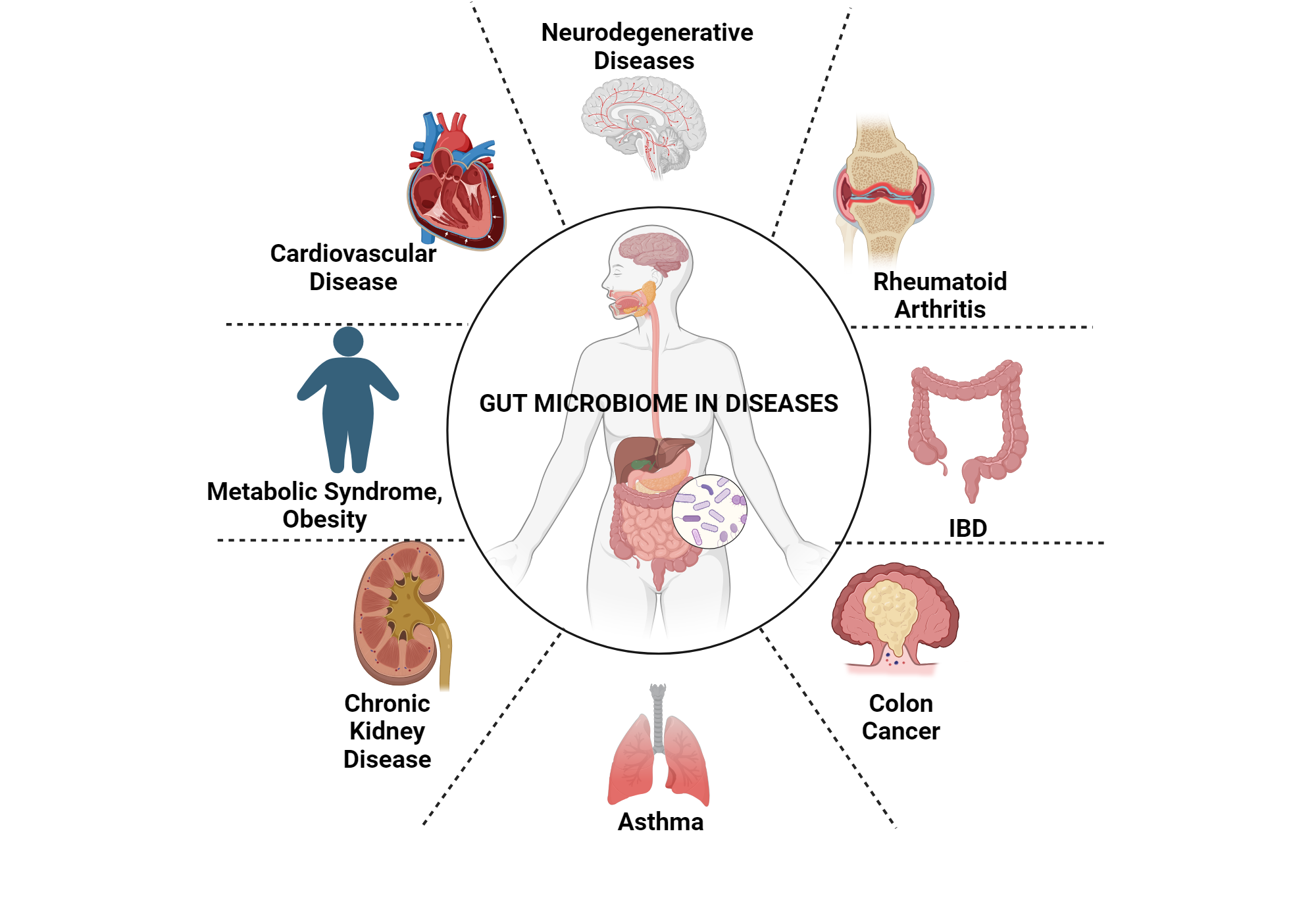
Over the past 5–10 years science has uncovered ever more evidence that the microbes living in the gut and the workings of our brain interact profoundly. Observational studies have shown that in numerous neurological and psychiatric conditions — for example anxiety, depression, autism spectrum disorder or Parkinson's disease — the gut flora of those affected differs from that of healthy people[4][5]. For a long time, however, it was uncertain whether the changes in the microbiome contribute to the development of these diseases or are merely a consequence of them. Among the known modes of the two-way communication between the brain and the gut are the neural pathways (such as the vagus nerve), hormonal and neurochemical signals (e.g. stress hormones or peptides produced in the gut), inflammatory messengers mediated by the immune system, and the molecules produced by the gut microbes themselves[3]. The latest research focuses precisely on uncovering these molecular and cellular mechanisms. We now know that gut bacteria produce numerous neurotransmitters, such as serotonin, dopamine and GABA, as well as short-chain fatty acids (SCFAs) and other metabolites, and even inflammatory cytokines, which can enter the circulation and reach the brain[3]. In addition, the composition of the microbiome also influences the activity and integrity of the enteric nervous system[6][7]. Overall, in the complex network of the gut–brain axis, the neural, hormonal, immunological and microbial components function closely interwoven.
One important lesson of the more recent results is that in certain cases there is a causal relationship between the processes taking place in the gut and the workings of the brain. In experimental models — mainly animal experiments, but by now in human studies too — it has been possible to show that modifying the gut flora can induce or prevent concrete brain changes and behavioural changes[8][9]. Let us look at the mechanisms behind this, and at the most exciting recent discoveries in gut–brain axis research.
One of the most important points of connection between the gut flora and the brain is the chemical substances produced by the microbes. These substances — whether metabolites, neurotransmitters or cell-wall components — can reach the brain via the bloodstream, or, acting locally, can trigger neural signals.
A remarkable example is the case of a recently identified bacterial metabolite, the molecule 4-ethylphenyl sulfate (4EPS). This compound is produced by gut bacteria during the breakdown of the amino acid tyrosine, and it had already been discovered that it is present in greater amounts in the blood of children with autism spectrum disorder[10]. Brittany Needham and colleagues in 2022 (Caltech; Nature, 2022) demonstrated in a mouse experiment that 4EPS is able to cross the blood–brain barrier and has a dramatic effect on the brain[11][12]. In mice whose gut flora produced large amounts of 4EPS, anxiety-like behaviour developed: placed in a new environment, they moved less and tended to hide, as if a predator were lurking[13][14]. Brain imaging showed increased activity in the centres of fear and anxiety, and the researchers found that 4EPS disrupts the functioning of the oligodendrocytes[12]. These cells produce the myelin sheath, the insulating layer that coats the axons (nerve fibres), just as plastic insulation coats cables — and they are crucial in the rapid transmission of signals between nerve cells. Under the effect of 4EPS, the oligodendrocytes in the mice's brains remained more immature and a thinner myelin sheath formed on the axons[12]. In essence, the molecule released by the microbiome slowed down the development of the insulation of the neural "cables", which appeared at the level of behaviour in the form of anxiety.
It is extremely exciting that this process could also be reversed. Needham and colleagues gave the 4EPS-exposed mice an experimental drug that stimulates myelin production. The result: normal myelin-sheath formation was restored in the mice, and the anxious behaviour also eased[15]. That is, a brain change caused by a microbial molecule — thinner axon sheaths and thus functional disturbance — could be reversed pharmacologically, precisely by counteracting the microbiome's effect in the brain. The study's title aptly put it: "a gut-derived metabolite alters brain activity and anxiety behaviour in mice"[16]. This is one of the first direct pieces of evidence that a concrete bacterial metabolite can establish a connection between the gut and the brain and modulate complex behaviour.
Another important route of the gut–brain axis leads through the immune system and inflammatory processes. In recent years chronic inflammation has come to the fore in the explanation of numerous psychiatric and neurological diseases — for example, some subtypes of depression have also been shown to be associated with a higher level of inflammation in the body (e.g. the level of cytokines such as IL-6). But how can the gut cause inflammation, and how does this affect the brain? An excellent example of this is a very recent discovery. Bang and colleagues in 2025 (Harvard Medical School, J. Am. Chem. Soc., DOI:10.1021/jacs.4c15158) found a bacterium — Morganella morganii — that produces unusual lipid molecules in the gut[17]. These lipids resemble the cardiolipins found in our own cells, but a compound considered an industrial pollutant, diethanolamine (DEA), is built into their structure[18]. The researchers showed that these "chimera-like" phospholipids of microbial origin trigger a powerful immune reaction: they activate the TLR2/TLR1 receptors of the innate immune system, following which large amounts of the aforementioned inflammatory cytokine, IL-6, are released[19]. Whereas normal cardiolipins are characterised by a weaker immune response, the administration of the abnormal lipid produced by Morganella induced, under experimental conditions, a stormy inflammatory response — known in the field as a cytokine storm.[19][20]
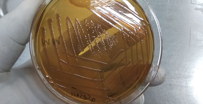
Morganella morganii
What is the significance of this from the point of view of the brain? Well, the multi-front study by Bang et al. also showed that Morganella morganii is often overgrown in the gut of depressed patients, and their blood IL-6 level was also elevated[21][22]. This suggests that a member of the gut microbiome can contribute to the development of depression via an inflammatory route — at least in certain people, under certain environmental conditions. The pollutant called DEA is, incidentally, a compound used in numerous industrially produced products, so the discovery also sheds light on how environmental pollution (micropollutants) can affect mental health through the gut bacteria[23]. According to the Harvard researchers, this "faulty" bacterial product could even serve as a biomarker for recognising a subgroup of depression, and, more importantly, it offers a new therapeutic target[24][25]. For if the inflammatory lipids of the gut bacteria cause the disease, then in principle treatments could be developed that neutralise these molecules or inhibit their formation. Moreover, the study's authors suggest that certain forms of major depression could even be understood as an autoimmune inflammatory disease that might also respond to immunomodulatory drugs[26][27]. Naturally, further research is needed to establish for how large a proportion of depression cases this mechanism is responsible, and how it fits into the complex picture of the disease[25]. In any case, this discovery clearly demonstrates that an immune activation triggered by a gut bacterium can influence the workings of the brain and mood, linking the microbiome to mental illnesses.
The third pillar of the gut–brain axis is the neural connections themselves. The main nerve running between the central nervous system and the gut, the vagus nerve, functions as a kind of information superhighway: it carries messages in both directions. The brain's signals affect gut function (just think how, in a stressful situation, digestion can speed up or come to a halt), but signals also arrive from the gut towards the brain. The enteric nervous system (ENS) — the independent neuron network in the wall of the gut — is not merely an executor of the brain's commands but pre-processes the sensory information coming from the gut, and even has its own reflex circuits. This is why the ENS is called the "gut's brain". An important realisation is that the state of the enteric nervous system is influenced by the gut flora: Fernando A. Vicentini and colleagues in 2021 (University of Calgary, Microbiome journal) showed that in healthy mice, wiping out the gut bacteria with antibiotics leads to the mass death of enteric nerve cells[28][29]. In the experiment, in the gut of adult mice treated with an antibiotic cocktail the number of neurons decreased drastically in the small and large intestine, and moreover the amount of glial cells (supporting nerve cells) also diminished in the ileum[28]. In parallel, their gut function was also disturbed: gut motility slowed, the permeability of the intestinal wall increased, and other functional abnormalities appeared[30]. All this suggests that the presence of a normal microbiome is essential for maintaining the gut's neural network. However, when the researchers stopped administering the antibiotic, the animals' gut flora eventually recovered spontaneously, and together with it new nerve cells formed in the gut — that is, the process of enteric neurogenesis was set in motion[29][31]. Within a few weeks the number of enteric neurons and glial cells rose to the normal level, and the gut-function disturbances ceased. Further experiments also revealed the underlying mechanisms: it turned out that a few key molecules among the gut bacteria are responsible for the survival and regeneration of the neurons. In the presence of lipopolysaccharide (LPS) — a molecule found in the cell wall of certain bacteria — fewer neurons died during the antibiotic course, while by supplementing the short-chain fatty acids (SCFAs) produced by the bacteria it was possible to "regrow" the lost nerve cells in the mice's gut[31][32]. That is, LPS exerted a protective effect on the neurons (presumably giving a "survival" signal via the immune system's TLR4 receptors), while acetic acid, butyric acid and other short fatty acids stimulated the division and differentiation of the neural stem cells, promoting the formation of new neurons[31][33]. This work pointed out that the gut flora actively maintains the gut's neural network, and if this support ceases (e.g. in a germ-free state or under the effect of antibiotics), degenerative processes begin in the ENS, which also affects the entire gut–brain communication[34][35].
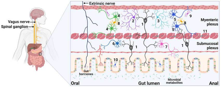
Regarding the neural connections, we must also mention the role of the vagus nerve. The vagus runs from the brainstem to the abdominal organs, and its sensory branches gather information from many points of the gut and transmit it towards the brain. For example, certain hormones produced in the gut (such as peptides that induce a feeling of satiety) or neurotransmitters released by the microbes can signal to the brain via the vagus nerve. According to one extreme but all the more interesting hypothesis, some neurodegenerative diseases may originate in the gut and spread by a neural route towards the brain. In connection with the pathological protein aggregates observed in Parkinson's disease (the so-called alpha-synuclein plaques), it has been suggested that they may form first in the nerve cells of the gut and from there "travel up" along the fibres of the vagus nerve into the brainstem and then on to other areas of the brain[36]. This is supported by some animal-experiment and human observations — for example, one study showed that when the pathological gut flora from a Parkinson's patient was transplanted into mice, it worsened the Parkinson-like symptoms and brain changes in the mice[37]. Moreover, in people whose vagus nerve had been surgically cut years earlier for some reason, Parkinson's disease developed less often — which indirectly suggests that part of the disease may indeed spread via the vagus. Although this area requires further research, the essential point is: in the gut–brain axis, the nerves also physically connect the two organs, and certain signals (even pathological proteins) can be transported along them.
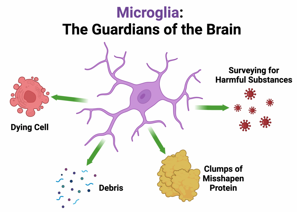
The boundaries of the immune system and the nervous system blur in the microglia cells — these are the brain's special immune cells, which continuously monitor the environment of the nervous tissue and, if necessary, intervene (clearing away debris, destroying pathogens, aiding the regeneration of nerve cells, but also potentially causing inflammation). Not surprisingly, the state of the microglia is also shaped by the gut flora. Huang et al. in 2023 (Third Military Medical University, China; Molecular Psychiatry) showed through experiments on mice that in the brain of germ-free (bacteria-free) animals the gene-activity pattern of the microglia differs radically from that of normal (SPF – specific pathogen free) mice[38]. Put more simply: in the absence of gut bacteria, the functioning of the brain's immune cells is re-tuned. The changes were moreover different from one brain area to another: a different gene network was activated in the prefrontal cortex and in the hippocampus, but the common denominator was that the change mainly affected the microglia cells[39][38]. Certain microglia subtypes appeared in greater proportions in the germ-free mice, while other types disappeared — and these changes were reversible when the researchers "re-established" the normal gut microbes in the animals[40][38]. That is, the microglia cells respond flexibly to the presence or absence of the peripheral microbiome. Why is this interesting? Huang et al. also noticed that the gene-expression patterns of the microglia that developed under bacteria-free conditions strikingly resemble the changes seen in the brains of human Alzheimer's and major depressive patients[41][38]. In addition, they found corresponding changes in the mice's behaviour: the germ-free mice showed less "hopelessness" in a swimming test (they did not give up swimming within 5 minutes) and performed better cognitively (memory) in certain tasks — which the re-establishment of the normal flora changed again[42][43]. These results suggest that the microbiome fine-tunes the functioning of the brain's immune cells, and that disturbances of this may contribute to diseases such as Alzheimer's or depression[44][38]. For example, it is possible that, due to the absence or presence of certain gut bacteria, the microglia become excessively pro-inflammatory or, conversely, fail to perform their protective functions properly — and this can, in the long term, promote neurodegenerative processes. In the future, the targeted influencing of such mechanisms (e.g. modulating the communication between the microglia and the gut bacteria) could represent a new therapeutic direction in brain diseases[45][46].
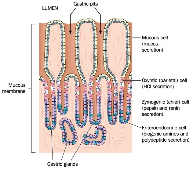
The gut–brain axis affects the brain not only through the nervous and immune systems in the classical sense, but also via hormonal regulation. In our gut there are special sensing and hormone-producing cells, the so-called enteroendocrine cells (EECs). These cells continuously monitor the composition of the gut contents and, depending on this, release hormones that influence digestion, appetite and numerous metabolic processes. The hormones also reach certain areas of the brain via the bloodstream, signalling for example satiety or hunger. Tan and colleagues in 2024 (UT Southwestern Medical Center, Nature Metabolism) shed light on how closely the functioning of the EECs is interwoven with the gut microbiota[47]. They developed a genetic mouse model in which the enteroendocrine cells of the colon are missing. These EEC-deficient mice, surprisingly, began to eat greedily, became obese and developed metabolic disturbances[48]. When the causes were examined, it turned out that the composition of their gut flora differed drastically from normal, and a certain metabolite had accumulated in their faeces: glutamate, that is, an amino acid (the salt of the messenger glutamic acid)[49][50]. The researchers' elegant experiments proved that these microbiome changes are responsible for the obesity: if the bacterial flora of the EEC-deficient mice was wiped out with antibiotics, the overeating and obesity ceased; and if the gut flora of an EEC-deficient mouse was transplanted into a germ-free (initially bacteria-free) mouse, the latter too began to put on weight[51]. That is, the changes in the gut flora were necessary and sufficient for the metabolic effects. Further metabolomic analyses revealed that in EEC deficiency certain bacterial groups began excessive glutamate production in the colon[52]. Glutamate — besides being an important neurotransmitter — can be one of the stimulators of the vagal sensory nerves in the brainstem: it increases the activity of the appetite centre. Moreover, glutamate delivered experimentally directly into the colon also increased food intake in the animals[52][50]. From this study a completely new gut–brain signalling pathway emerges: the interaction between the colon's hormone-producing cells and the microbiome is able to regulate the energy balance of the whole organism. If the hormonal "switch" (the EEC) drops out, the balance of the gut flora is upset (dysbiosis develops), and the extra microbial glutamate released in this way conveys the message "eat more!" towards the brain. This realisation sheds light not only on a new mechanism of obesity and overeating, but also on the fact that the gut–brain axis can influence, beyond mood disorders, metabolism and appetite as well.
Knowing the above mechanisms, the question arises: how can these scientific results be applied in medicine, and how might they explain the origin of certain diseases? First, there is more and more evidence that in numerous neurological and psychiatric conditions a disturbance of the gut–brain axis plays a role. Such is, for example, depression, where — as we have seen — in some cases the inflammatory components of the gut flora may contribute to the symptoms[22][26]. The clinical consequence of this realisation is that, in the treatment of depression, it may be worth also looking towards reducing the activity of the immune system and the degree of inflammation (for example through anti-inflammatory therapies or the modulation of the gut flora). Some researchers are already experimenting with agents that bind or inhibit the cardiolipin-like molecules produced by Morganella — this could potentially be a new antidepressant strategy in the future.
In the case of anxiety disorders, microbiome research likewise opens new horizons. Following the study focused on the aforementioned 4EPS molecule, a biotech company (Axial Therapeutics) developed an experimental agent designated AB-2004, which is essentially an oral adsorbent — it binds aromatic molecules similar to 4EPS in the gut before they can be absorbed. This agent was tested on adolescents with autism spectrum disorder in an open clinical trial. Sarkis Mazmanian and colleagues in 2022 (Caltech/UCLA, Nature Medicine) reported that AB-2004 can be used safely, and that the levels of several bacteria-derived metabolites decreased significantly in the patients' blood and urine[9]. Moreover, after 8 weeks of treatment, an improvement appeared in several behavioural symptoms, particularly in the area of anxiety and irritability, as well as in the easing of gastrointestinal complaints (though, in the absence of a control group, the results must be evaluated with caution)[9]. This is one of the first swallows in the direction of trying to treat psychiatric abnormalities — in this case certain symptoms of autism — by intervening in the gut. Similar experiments are under way with faecal transplantation (FMT) too. In any case, the targeted manipulation of the gut flora (be it bacterial removal, supplementation or metabolite binding) as a therapeutic strategy is no longer science fiction today but an actively researched field.
In connection with neurodegenerative diseases — such as Parkinson's disease or even Alzheimer's disease — there are also more and more indications that the gut–brain axis participates in the processes of the diseases' development. Faecal-microbiome analyses of Parkinson's patients consistently show dysbiosis (an altered bacterial composition), and — as already mentioned — some symptoms of Parkinson's can be transferred in animals via bacteria[37]. Hyunji Park and colleagues in 2025 (KAIST, South Korea; Nature Communications) identified one concrete culprit: the bacterium Streptococcus mutans, which overgrows in the gut of Parkinson's patients[53]. This pathogen, also responsible for tooth decay, normally lives in the mouth, but it seems it also survives when it reaches the gut, and produces an enzyme called UrdA, which creates a molecule called imidazole propionate (ImP) from the amino acid histidine[53][54]. Park et al. showed that the level of ImP is high in the blood and in the cerebrospinal fluid of Parkinson's patients, and that the molecule is able to cross the blood–brain barrier. What is more, they colonised mice with this bacterium (as well as with an E. coli strain carrying the gene of the UrdA enzyme), and found that in the animals ImP accumulated in the brain, which was followed by the appearance of Parkinson-like symptoms: in their brains the number of dopamine-producing nerve cells decreased, the glial cells (the brain's immune cells) were activated, and movement disorders appeared[54][55]. Furthermore, when pure ImP molecule was given to the mice, it itself induced the characteristic pathological symptoms of Parkinson's, precisely by activating the mTORC1 signalling pathway in the brain[55]. By inhibiting mTORC1 (with an agent called rapamycin), however, the nerve-cell death and movement disorders caused by ImP could be prevented[56][57]. According to the work's conclusion, ImP is a microbiome-derived toxic molecule that may directly contribute to the pathogenesis of Parkinson's disease and could be a new therapeutic target in treating the disease[58]. This is a huge step forward, since it provides concrete evidence for a causal chain: bacterium → metabolite → brain damage → disease. It also raises the possibility that in the future Parkinson's disease might be slowed in part by influencing the gut flora (e.g. by wiping out Streptococcus mutans or neutralising ImP).
Another study, also from 2025 (Morais et al., Caltech, npj Parkinson's Disease[7]), pointed out that the normal gut flora can aggravate Parkinson's disease by increasing the activity of the mitochondria in the brain and thereby the oxidative stress. The researchers compared Parkinson-model mice (genetically modified mice that overproduce the human alpha-synuclein protein and are thus prone to Parkinson-like symptoms) such that one group had a normal gut flora and the other group was germ-free. They found that in the brains of the mice with a normal microbiome, in the striatum area the mitochondria became hyperactive, and as a result a great many free radicals and peroxide derivatives were produced — that is, the cells suffered oxidative damage[59]. In contrast, the brains of the germ-free (GF) mice were more protected: in them antioxidant proteins that neutralise the harmful free radicals were expressed at a higher level[60]. Even more convincing evidence came when the mice with an intact microbiome were given an antioxidant drug, which improved their motor symptoms, while if the antioxidant defence mechanisms were artificially inhibited in the germ-free mice, their movement worsened (but only in those in which the human pathological alpha-synuclein protein was present)[61][62]. From this it follows that the gut flora contributes to the symptoms of Parkinson's by creating a harmful environment burdened with oxidative stress in the brain, while in its absence the brain defends itself better against the disease[60][62]. This realisation is important because it highlights that in Parkinson's disease too it may be worth targeting the gut flora — for example with pre- or probiotics that reduce the proportion of oxidative-stress-inducing bacteria, or indeed increase that of the protective ones.
Of course, not every disease can be traced back to the microbiome, and we must not oversimplify the picture. In most cases the gut–brain axis is only one factor among many that contribute to the condition. In the case of Alzheimer's disease, for example, although indications of a possible role of the gut flora have been found (e.g. in connection with microglia transformation, see above), among the main drivers of the disease there remain genetic predisposing factors and other processes taking place in the brain. At the same time, a new outlook is beginning to take shape in medicine: some mental and neurological diseases are now interpreted at the systems level, in the whole body–brain interaction, rather than as isolated disorders of the brain. In this framework the gut, as a central "traffic hub", receives special attention.
Research on the gut–brain axis is currently developing explosively, but there are still many open questions. In the future the emphasis will probably be on:
In summary, research on the gut–brain axis is an exciting, multidisciplinary field that is fundamentally changing our conception of the relationship between brain and body. It teaches us that the human body is an integrated ecosystem, in which the brain, the gut and the microbes living within us are all important "inhabitants" and regulators of this ecosystem.
Twenty-first-century medicine is beginning to realise that our health is determined by the unity of our brain and body — indeed, by the superorganism as a whole that we form together with the microorganisms living within us. Research on the gut–brain axis is still in its infancy, but it already allows insight into how closely our thoughts and feelings are tied to the tiny beings living within us and to the signals of our internal organs. In the words of Yuval Noah Harari, the human being is, instead of "animals and machines", now a blend of animals, machines and microbes. As the journalist Ed Yong wrote, "we are not alone in our own minds" — our gut bacteria whisper to us too. The research of the coming years may make it possible for us to learn to "converse" with this invisible organ of ours, the microbiome, and thereby to heal both body and soul along new paths. Understanding the gut–brain axis ultimately brings us closer to treating the human being as a complete, holistic system — for part of our being is also what lives within us.[22][18]
We are happy to answer your professional questions — we respond to every enquiry.
Write to us